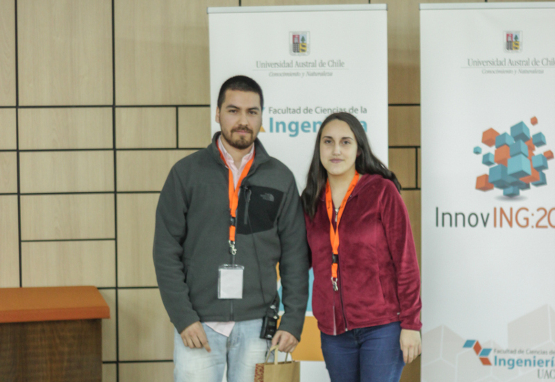
The ex-student of the Medical Images Analysis Group, Ignacio Espinoza, obtained First Place in the paper contest, with his work “Virtual reality software for the exploration of the brain in 3D“. The app allows to visualize lobes, convolutions, subcortical structures and the main fiber fascicles of the brain. It was made using Unity and Blender software, and it allows interaction via the Oculus Rift virtual reality lens and the Leap Motion device.

Felipe Silva (who is currently working on his thesis) obtained the Second Place, with his work “Color detector device for people with visual disability”.
The device uses a RGB color sensor capable of detecting colors, where two modes were implemented: the first one reproduces the name of the color through a speaker, whereas the second one uses a combination of three semitones, allowing the relation of colors and music without depending on a particular language. This device aims to improve the life quality of a blind person in daily chores like, for example, knowing the color of the clothes they wear.

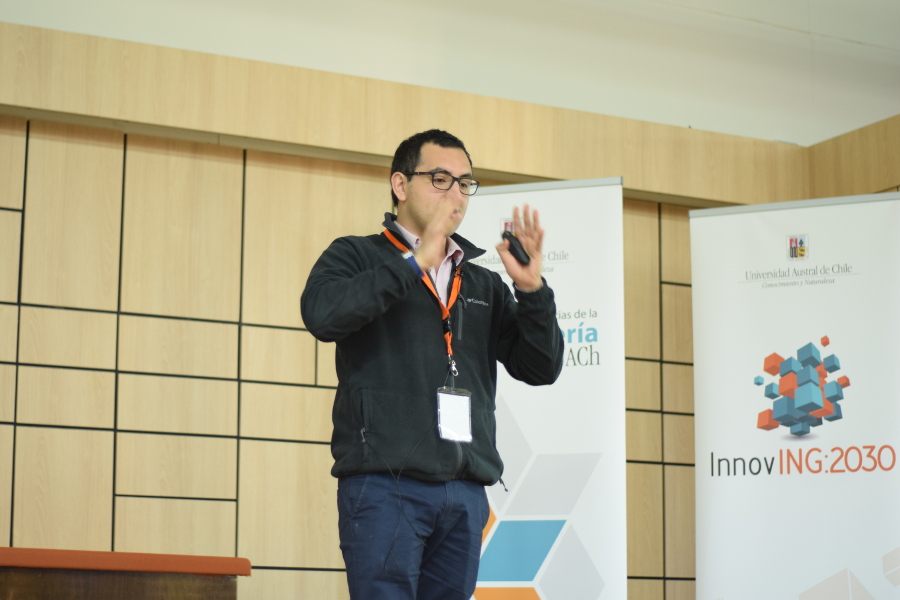
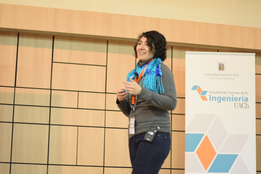
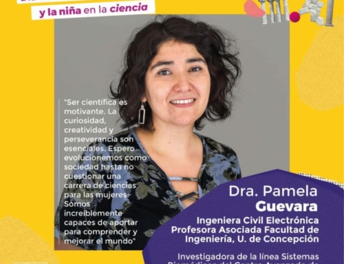
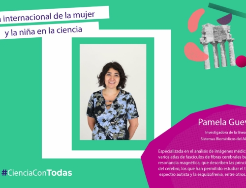
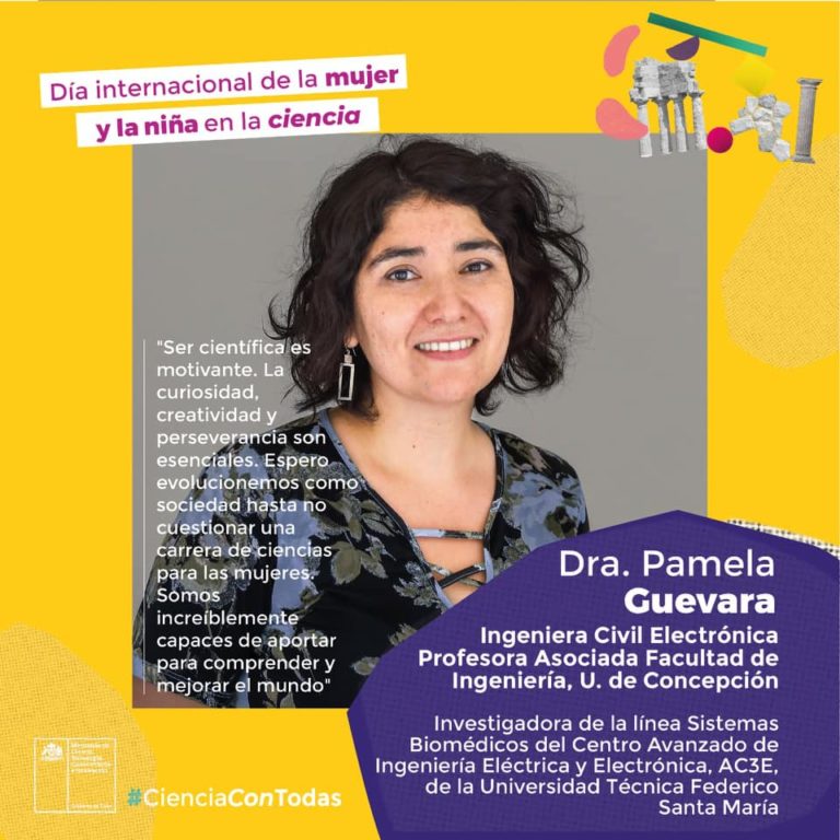
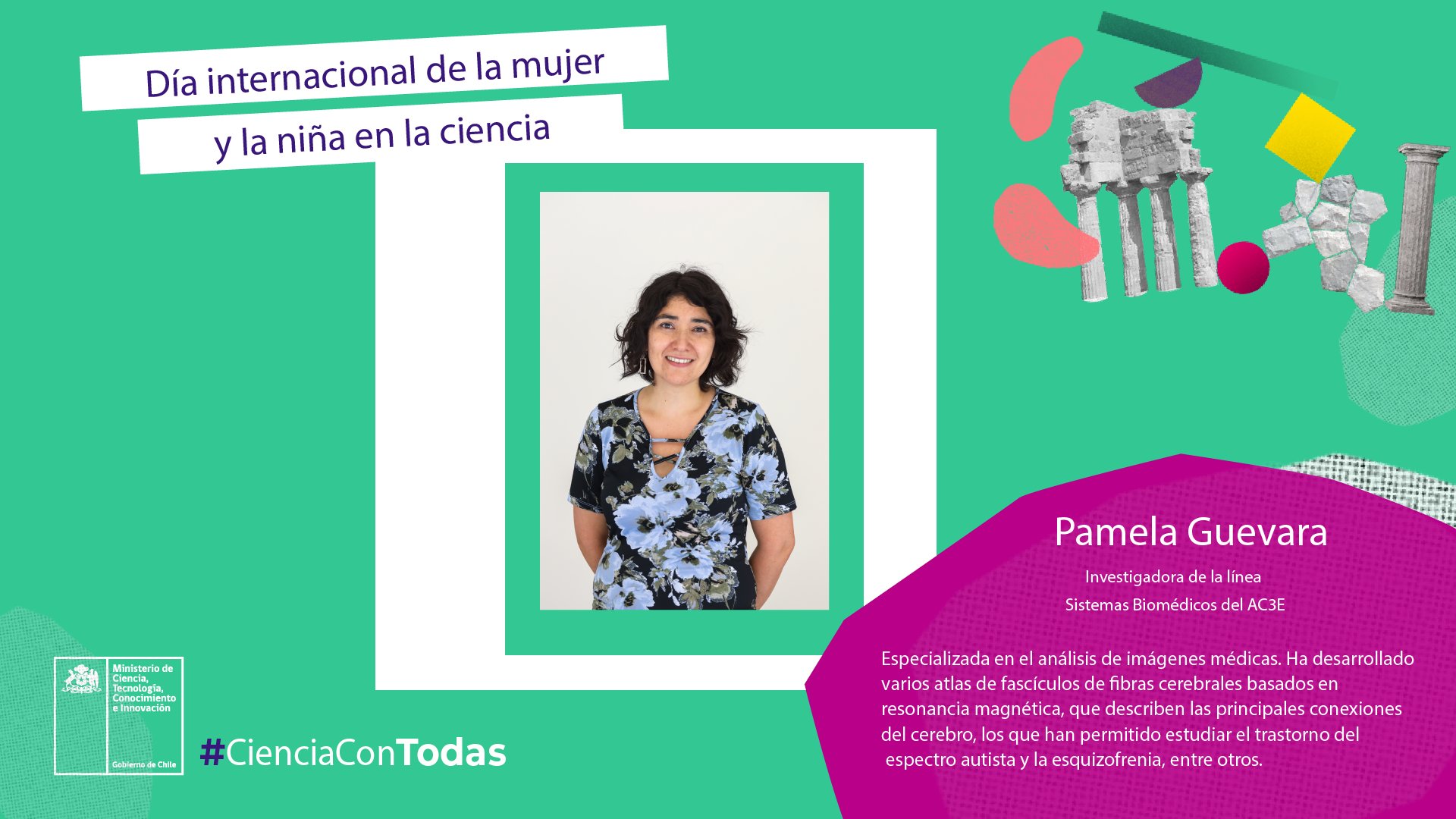
Professor Pamela Guevara dictated the presentation “Study of brain connectivity from difussion Magnetic Resonance”, in which she exposed the acquisition and preprocessing of dMR data.
Afterwards, she presented the main works developed by the Medical Images Analysis Group, including the creation of a brain fascicles atlas, and algorithms for the clustering of brain fibers and segmentation of fiber fascicles (all carried out in the context of the “FONDECYT 116127” and “Anillo ACT172121” projects).


Leave A Comment